
By ProHobby™ | Ecological Systems Authority
Lymphocystis is the aquarium disease that most commonly receives the wrong treatment for the wrong diagnosis — and where the correct response (no medication, optimise conditions, wait) is the most counterintuitive thing a hobbyist can do when confronted with visible, alarming growths on a fish they care about. The cauliflower-like white or pinkish nodules on fins and body surface look like something that should be treated aggressively. They should not be. Lymphocystis is a viral disease that is self-limiting in fish with good immune function — the fish’s own immune system resolves it over weeks to months, provided the environmental conditions that suppressed immunity in the first place are corrected. No antifungal, no antibiotic, no commercial disease treatment has any useful activity against an iridovirus. Reaching for medication is not just unhelpful; it adds pharmaceutical stress to a fish that needs the opposite.
Understanding why Lymphocystis looks the way it does, why no medication works, and what actually helps is the complete management picture for this disease.
Table of Contents
- What Lymphocystis Is — The Iridovirus Biology
- Why the Growths Look the Way They Do — The Hypertrophied Fibroblast
- Symptoms — Identifying Lymphocystis Correctly
- Differential Diagnosis — The Conditions Lymphocystis Is Mistaken For
- Why There Is No Medication — And Why This Is Correct
- What Actually Helps — Management Without Treatment
- Secondary Bacterial Infection — The One Treatable Complication
- Surgical Removal — When It Helps and When It Doesn’t
- Transmission and Biosecurity — How Lymphocystis Spreads
- Species Most Susceptible
- Lymphocystis in Marine Fish
- Prognosis — The Honest Timeline
- India and Delhi NCR — Specific Considerations
- Frequently Asked Questions
1. What Lymphocystis Is — The Iridovirus Biology
Lymphocystis is caused by Lymphocystivirus — a member of the family Iridoviridae, a family of large double-stranded DNA viruses that infect ectothermic animals (fish, amphibians, reptiles, and invertebrates). The specific virus affecting aquarium fish — historically referred to as Lymphocystis disease virus (LCDV) — infects connective tissue cells called fibroblasts and produces the most visually dramatic pathological effect of any common aquarium disease: individual cells enlarged to dimensions visible to the naked eye.
Iridoviruses are notably different from the bacterial and parasitic pathogens that cause most other aquarium diseases in one critical way: they are not susceptible to any antibiotic, antifungal, antiparasitic, or other pharmacological agent currently available to aquarium hobbyists or veterinarians. Viral diseases do not respond to antibiotics — this is true in human medicine, in veterinary medicine, and in fish medicine equally. The virus must be cleared by the immune system or not at all.
The Lymphocystivirus is also a latent pathogen in many fish populations — meaning the virus may be present in a fish without producing any visible disease, held in check by normal immune function. This latent state is the reason Lymphocystis frequently appears after a stress event that depresses immunity — the virus was already present, and the immune suppression allowed it to establish active infection. The Science of Fish Stress covers the cortisol-immunity cascade in detail.
2. Why the Growths Look the Way They Do — The Hypertrophied Fibroblast
This is the biology that no hobbyist guide explains, and understanding it immediately makes the visual presentation intuitive.
When Lymphocystivirus infects a fibroblast cell, it essentially commandeers the cell’s machinery to replicate viral particles while simultaneously preventing normal cell division and death. The infected cell does not divide, does not die on schedule, and does not shed — it simply grows. And grows, and grows. An infected fibroblast cell can reach 100,000 times its normal volume over the weeks of active viral replication — producing a single cell visible to the naked eye, which is a genuinely extraordinary biological event. Most animal cells are 10–30 micrometres in diameter; a hypertrophied Lymphocystis cell can reach several millimetres.
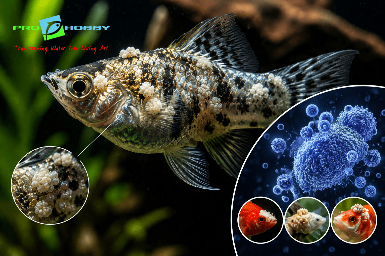
What is visible on the fish as a “nodule” or “growth” is typically a cluster of these massively enlarged individual cells, each surrounded by its own thick hyaline membrane that the cell produces in response to viral activity. Under a hand lens or low-power microscope, the individual cell structure is clearly visible — a feature that makes Lymphocystis immediately diagnosable by anyone willing to look closely, distinguishing it from bacterial plaques, fungal growth, and parasitic cysts which all have different structural appearances.
The nodules are not tumours. They are not cancerous. They are individual infected cells that have become pathologically enlarged. When the immune system ultimately clears the viral infection from those cells, the enlarged cells undergo normal apoptosis (programmed cell death) and the growths disappear without leaving structural damage to the underlying tissue in most cases.
3. Symptoms — Identifying Lymphocystis Correctly
The characteristic appearance: White, off-white, pinkish, or grey-white nodules on the fin edges, fin membrane, and body surface. The nodules have a distinctive texture — irregular, slightly rough, sometimes described as resembling cauliflower or raspberry in structure. They may occur singly, in small clusters, or in larger aggregations covering significant fin area.
Location pattern: Lymphocystis most commonly appears on fin edges — the tips of dorsal, caudal, and anal fins — and on the body surface near fin bases. Oral Lymphocystis (growths at or near the mouth) occurs and is particularly significant because it can interfere with feeding. Internal Lymphocystis affecting visceral organs occurs, diagnosed only post-mortem.
Size range: Individual nodules visible to the naked eye, from approximately 1mm to several millimetres in diameter. Clusters of nodules can cover centimetre-scale areas of fin membrane.
Fish behaviour: Typically minimal change in the early stages. Most fish with Lymphocystis continue eating and behaving relatively normally — the nodules are not acutely painful in the way that parasitic or deep bacterial infections are. Behavioural changes (reduced appetite, lethargy) occur when growths become extensive, when oral involvement interferes with feeding, or when secondary bacterial infection develops.
Progression timeline: Nodules typically first appear and enlarge over 2–6 weeks, plateau in size, and then gradually regress over 4–12 weeks as immune response mounts. The total visible disease course in a fish with good water quality and resolved stress is often 2–4 months from first appearance to complete resolution.
4. Differential Diagnosis — The Conditions Lymphocystis Is Mistaken For
This table is the most important clinical tool in Lymphocystis management because misdiagnosis leads directly to wrong treatment.
| Feature | Lymphocystis | Columnaris (bacterial) | Saprolegnia (fungal) | Ich |
|---|---|---|---|---|
| Appearance | Irregular nodules, cauliflower texture, discrete lumps | Flat white-grey attached patches, cottony or eroding | Three-dimensional fluffy tufts at wound sites | Discrete 1mm white spots uniformly distributed |
| Texture | Rough, nodular, structural | Flat, surface-attached | Fluffy, extending into water | Smooth, salt-grain appearance |
| Location | Fin edges, fin membrane, body | Any body area, fins, mouth | Specifically at wound sites | Across body and all fins |
| Progression | Slow — weeks | Fast — hours to days | Moderate | Moderate |
| Response to antibiotic | None | Effective | None | None |
| Response to antifungal | None | None | Effective | None |
| Response to ich treatment | None | None | None | Effective |
| Correct response | Water quality, no medication | Gram-negative antibiotics | Antifungal + address primary wound cause | Heat, salt, medication |
Key diagnostic rule: If the white growth is three-dimensional, nodular, irregular in texture, and on fin edges or the body surface without a specific wound site as origin, it is Lymphocystis. If it does not respond to any antibiotic or antifungal treatment over 5–7 days, Lymphocystis should be reconsidered as the diagnosis. For definitive identification: examine a nodule under a hand lens or low-power microscope — the individual enlarged cells with their thick hyaline membranes are distinctly visible and unlike any bacterial or fungal growth pattern.
Warts and cysts: Some fish develop benign epidermal papillomas (warts) that superficially resemble Lymphocystis nodules but are composed of normal epidermal cells rather than hypertrophied fibroblasts. Fish cysts from various causes also produce visible lumps. All of these require microscopic examination for confident differentiation. The External Protozoan Parasites guide covers additional differential considerations for surface changes in fish.
5. Why There Is No Medication — And Why This Is Correct
This section exists because the absence of a treatment recommendation needs justification — hobbyists are conditioned to expect that every fish disease has a product or a protocol.
Viruses are not living organisms in the conventional sense — they are nucleic acid packages that hijack host cell machinery to reproduce. The mechanisms that antibiotics exploit (bacterial cell walls, protein synthesis pathways, DNA replication enzymes specific to bacteria) do not exist in viruses. Antifungals target fungal cell membrane components (ergosterol) that are absent in viral particles. Antiparasitics target the neuromuscular systems of multicellular parasites. None of these mechanisms have any point of action in an iridovirus.
There are no licensed antiviral medications for fish in any market. Experimental use of antiviral compounds from human or veterinary medicine has not produced meaningful results against Lymphocystivirus in aquarium fish. Treating a Lymphocystis case with antibiotics, antifungals, or antiparasitic agents is guaranteed to produce no improvement in the viral condition while adding pharmaceutical stress to the fish and potentially disrupting the biological filtration that maintains water quality. Quarantine vs Medication.
The immune system of a fish in excellent water quality is a more effective Lymphocystis treatment than any product on a shelf. The management goal is to remove everything that suppresses it and let it do its work.
6. What Actually Helps — Management Without Medication
Step 1: Identify and correct the stressor that allowed active infection to develop.
Lymphocystis typically becomes clinically visible after an immune-suppressing event. The most common triggers in aquarium fish: water quality deterioration (ammonia, nitrite spikes, KH depletion, temperature swings), chronic overcrowding stress, recent transport, aggressive tank mate encounters, or sustained poor nutrition. Identifying and eliminating the specific trigger is the first and most important management step. Test ammonia, nitrite, pH, and temperature. Review stocking density. Complete Water Chemistry Guide.
Step 2: Optimise water quality to the upper end of the species’ acceptable range.
This means not just acceptable parameters but genuinely excellent ones — zero ammonia, zero nitrite, nitrate below 10–15 ppm, stable pH with KH above 4 dKH, temperature stable at the species’ ideal (not merely tolerated) level. Regular water changes using the Water Change Calculator. How to Do a Water Change.
Step 3: Improve nutrition.
Vitamin C specifically is associated with immune function in fish and is depleted rapidly in processed foods stored at room temperature. Supplementing food with vitamin C (ascorbic acid solution applied to pellets before feeding), adding variety with fresh or frozen foods, and ensuring the fish is actually consuming adequate nutrition — particularly relevant if oral Lymphocystis is reducing feeding — all support immune recovery.
Step 4: Reduce stress from all sources.
Minimise disturbance, manage aggressive tank mates, ensure appropriate hiding structures and territory, and consider whether the tank is appropriately sized and decorated for the species. The immune suppression of chronic social stress is real and measurable. Isolate the fish in a hospital tank if tank mate aggression is occurring or if the Lymphocystis is extensive enough to make the fish a target.
Step 5: Wait.
Weeks. Possibly months. Lymphocystis resolution is slow. Nodules that are not growing and a fish that is maintaining normal behaviour in excellent water quality are a treatment success, even if the nodules have not yet visibly reduced. Patience is part of the management protocol.
7. Secondary Bacterial Infection — The One Treatable Complication
While Lymphocystis itself cannot be treated, the enlarged, disrupted cells at the lesion surface are vulnerable to bacterial colonisation — particularly by the opportunistic gram-negative bacteria responsible for fin rot and scale rot.
Signs of secondary bacterial infection at a Lymphocystis site:
- Reddening or haemorrhaging at the margin of the Lymphocystis nodules
- Expanding erosion of fin tissue around the nodule cluster
- White-grey necrotic tissue developing around the growth
- Accelerating deterioration that does not fit the typical slow Lymphocystis timeline
Secondary bacterial infection at Lymphocystis sites is the appropriate indication for antibiotic treatment — gram-negative antibiotics (kanamycin sulphate or nitrofurazone) in a hospital tank following the fin rot treatment protocol. This treats the bacterial complication, not the viral disease itself, but preventing secondary infection from causing additional tissue damage supports the conditions for immune-mediated viral resolution.
8. Surgical Removal — When It Helps and When It Doesn’t
Lymphocystis nodules can be physically removed — the enlarged cell clusters are discrete enough to excise — but surgical removal is rarely the appropriate first response and carries meaningful risks.
When surgical removal may be appropriate:
- Oral Lymphocystis interfering severely with feeding, where the nodule is large enough to prevent the fish eating normally
- A very large single nodule on the body surface that is causing functional impairment
- Cases where the nodule is clearly becoming necrotic and secondary infection risk is high
The risks of surgical removal:
- The procedure itself requires brief anaesthesia (clove oil), clean instruments, and post-procedure wound management with diluted povidone-iodine — creating exactly the kind of physical wound that enables secondary bacterial infection
- Disrupting an intact Lymphocystis nodule releases large quantities of viral particles into the water, potentially increasing transmission risk to other fish
- In most cases, the immune system will resolve the nodule without surgical intervention given adequate time and optimal conditions
When surgical removal is not indicated:
- Small or moderate nodules on fin edges that are not causing functional impairment
- Fish that are eating normally and behaving normally despite visible growths
- Nodules that appear to be in the plateau or regression phase
If surgical removal is performed, isolate the fish in a hospital tank before and after the procedure, apply topical antiseptic to the wound site, and monitor closely for secondary bacterial infection in the days following. Septicaemia is a risk if bacteria enter the bloodstream through the wound.
9. Transmission and Biosecurity — How Lymphocystis Spreads
Lymphocystivirus spreads through direct contact with infected tissue and through water contaminated with viral particles from disrupted nodules.
Primary transmission routes:
- Direct physical contact between fish — fighting, fin nipping, or close proximity to a fish with actively shedding nodules
- Disruption of nodules (through aggression, handling, or surgical removal) releasing concentrated viral particles into shared water
- Contaminated equipment (nets, siphons, scissors) used on an infected fish and then introduced to a clean tank
The latent infection complication: Because the virus can exist in fish without producing visible disease, a fish that appears healthy may be a carrier capable of transmitting Lymphocystivirus to other fish that are then stressed enough to develop active infection. This makes apparent clearance of Lymphocystis from a tank population not equivalent to elimination of the virus from that population.
Biosecurity measures:
- Quarantine all new fish before introduction — Quarantine and Biosecurity in Aquariums
- Separate equipment (nets, siphons) for tanks with confirmed Lymphocystis cases
- Do not deliberately disrupt or abrade nodules
- Remove fish with Lymphocystis from community tanks where aggression is occurring — the combination of nodule disruption from fin nipping and stress from being targeted significantly worsens both the viral disease and transmission risk
10. Species Most Susceptible
Lymphocystis has been documented in a very wide range of freshwater and marine fish species — over 100 species from numerous families — but some groups show consistently higher clinical prevalence in the aquarium hobby.
Cichlids: One of the most commonly affected groups. Oscars, flowerhorns, discus, and angelfish all appear in clinical Lymphocystis reports with notable frequency. The stress of cichlid territorial dynamics — particularly in cichlid community tanks where social hierarchy stress is sustained — creates the immune suppression context for Lymphocystis expression. Oscar Fish Care. Flowerhorn Cichlid Care.
Labyrinthfish (bettas, gouramis): Frequent reports, particularly in bettas kept in suboptimal conditions — the same population whose stress-immunity relationship was discussed in the betta care context. Betta Fish Care Guide.
Gouramis: Both dwarf and larger gourami species show notable susceptibility, possibly related to their general sensitivity to stress.
Livebearers: Guppies and mollies less commonly than cichlids or labyrinthfish, but documented.
11. Lymphocystis in Marine Fish
Marine Lymphocystis presents essentially identically to freshwater cases — the same nodular cauliflower-like growths on fin edges and body surface — and is caused by related but distinct Lymphocystivirus strains adapted to marine fish species.
Clownfish, wrasses, tangs, and various other popular marine species are affected. Management is identical: no medication (the viral biology is the same), correction of the stressors (which in reef and marine contexts include salinity swings, insufficient flow, incompatible tank mates, and the complex stability demands of reef water chemistry covered in Reef Aquarium Ecology and Collapse), and immune support through excellent water quality.
The secondary bacterial infection risk in marine Lymphocystis is at least as significant as in freshwater cases — the same gram-negative opportunists exploit disrupted viral lesion sites in marine systems as in freshwater.
12. Prognosis — The Honest Timeline
For a fish in excellent water quality with the stress trigger identified and resolved: Very good. Lymphocystis resolves spontaneously in the majority of immunocompetent fish within 2–4 months of onset. Resolution is sometimes faster; occasional cases take longer. Recurrence is possible if the fish experiences another significant immune-suppressing event.
For a fish with ongoing water quality problems or unresolved stressors: Poor. Without immune support, the virus continues active replication. Nodules expand, secondary bacterial infection becomes more likely, and the fish deteriorates progressively.
For a fish with extensive oral involvement preventing normal feeding: Guarded. If the fish cannot feed, nutritional status declines, immune function falls further, and a downward spiral can develop. Surgical removal of oral nodules may be necessary to allow normal feeding to resume.
The critical honest point: Lymphocystis is not always mild. In a stressed, immunocompromised fish in poor water quality, it can progress to extensive body surface involvement, secondary septicaemia, and death. The self-limiting nature of the disease applies specifically to fish with adequate immune function in good environmental conditions — not universally.
13. India and Delhi NCR — Specific Considerations
Summer temperature stress: Delhi NCR summer conditions — ambient temperatures pushing tank water above 30°C in uncontrolled rooms — produce the immune suppression that triggers Lymphocystis expression from latent infection. Cases of Lymphocystis appearing in summer in previously healthy fish, with no other identifiable change, are a consistent pattern. Temperature management is both prevention and the first management step. Aquarium Water Temperature in Indian Summer.
Hard water and cichlid keeping: Delhi NCR’s hard tap water sits outside the ideal chemistry for many cichlid species commonly kept in the region — oscars, discus, and angelfish all originate from soft water systems. The chronic mild osmotic stress of incorrect water chemistry is a real immune suppressor, and Lymphocystis in cichlids in Delhi NCR is frequently associated with water chemistry management that is adequate but not optimal. Correcting toward species-appropriate parameters is part of the immune support strategy. Hard Water Aquariums in Delhi NCR.
Import stress and latent virus: Fish entering the Indian market through standard import chains experience significant transport stress that both suppresses immunity and can trigger latent Lymphocystivirus expression. Lymphocystis appearing within the first 4–6 weeks after acquiring a new fish — from any source — is frequently import-stress triggered. This is precisely why quarantine before main tank introduction is standard practice: Quarantine and Biosecurity in Aquariums.
Frequently Asked Questions
What are the white lumpy growths on my fish’s fins?
Almost certainly Lymphocystis — a viral disease producing characteristically nodular, cauliflower-textured growths on fin edges and body surface. Confirm by examining the growth under a hand lens: individual enlarged cells with thick hyaline membranes are visible and distinctive. Test water quality immediately — Lymphocystis typically appears when immune function is suppressed by water quality deterioration or stress.
Is Lymphocystis contagious to other fish?
It can be. The virus spreads through disrupted nodules releasing viral particles and through direct contact. However, transmission requires the receiving fish to also be immunocompromised enough for active infection to establish — healthy fish in excellent water quality may be exposed without developing disease. Remove fish with Lymphocystis from tanks where fin nipping or aggression is occurring; do not deliberately disrupt nodules.
Can Lymphocystis kill fish?
Yes, in the wrong conditions. The disease is self-limiting in immunocompetent fish with good water quality — in these fish, the immune system resolves it over weeks to months without treatment. In fish with ongoing stress, poor water quality, or compromised immunity, the virus continues progressing, secondary bacterial infections establish at the lesion sites, and the fish can die. The self-limiting reputation applies specifically to well-managed fish, not universally.
What is the treatment for Lymphocystis?
There is no medication that treats Lymphocystis — it is a viral disease and viruses do not respond to antibiotics, antifungals, or antiparasitics. The correct management is: identify and eliminate the stress trigger, optimise water quality to the upper end of the species’ acceptable range, improve nutrition with vitamin C supplementation, reduce all social and environmental stress, and wait for immune-mediated resolution. Secondary bacterial infections at lesion sites are treated with gram-negative antibiotics.
How long does Lymphocystis last?
Typically 2–4 months from first visible growths to complete resolution in a fish with good immune support and optimal water quality. Some cases resolve faster; some take longer. Recurrence is possible if the fish experiences another significant immune-suppressing event.
My fish had Lymphocystis, the nodules disappeared, and now they’re back — why?
Recurrence indicates either the original stress trigger was not fully resolved, or the fish has experienced a new immune-suppressing event. Lymphocystivirus may remain present as a latent infection after visible resolution — the nodules disappearing means the immune system controlled the active infection, not that the virus was eliminated from the fish’s body. Any subsequent significant stress can trigger re-expression.