By ProHobby™ | Ecological Systems Authority
Fungal infection in aquarium fish is over-diagnosed, under-treated when it does occur, and — most significantly — confused with a bacterial disease that looks identical but requires the opposite treatment. The pattern is consistent: a hobbyist sees a white growth on a fish, reaches for an antifungal, sees no improvement or active worsening, and concludes the treatment is not working. In the majority of cases, the treatment is not working because the condition is Columnaris — a gram-negative bacterial disease — not fungal infection at all. The correct diagnosis determines everything. Getting it right starts with understanding what genuine fungal infection actually looks like.
Table of Contents
- What Causes Aquarium Fungal Infections
- Why Fungal Infection Is Always Secondary
- What Fungal Infection Looks Like — The Definitive Visual Test
- Distinguishing Fungal From Bacterial — The Most Critical Differential
- Treatment — Step by Step
- Fungal Infections in Spawning Tanks and Eggs
- After Treatment — Addressing the Primary Cause
- India and Delhi NCR — Specific Considerations
- Frequently Asked Questions
1. What Causes Aquarium Fungal Infections
Saprolegnia and Achlya species are the primary aquarium fungal pathogens — technically oomycetes (water moulds) rather than true fungi, but treated identically in aquarium management. They are present in virtually all aquarium water at low levels and cause clinical infection only when given an entry point: a wound, an area of damaged tissue, or severely immunocompromised tissue.
The immunity framework behind why healthy fish do not develop fungal infections: The Science of Fish Stress.
2. Why Fungal Infection Is Always Secondary
This is the most important biological fact about aquarium fungal disease: a healthy fish with intact skin does not develop fungal infection regardless of how much Saprolegnia is in the water. Fungal infection is not random misfortune. It is the consequence of something else that compromised the fish or created damaged tissue first.
Primary causes that enable fungal infection:
- Physical wounds from handling, net damage, fin nipping, or collisions with décor
- Bacterial infection sites where skin integrity has been compromised by fin rot or body ulcers
- Anchor worm removal wound sites
- Unfertilised eggs in spawning tanks — these die first and rapidly develop fungal growth that spreads to viable adjacent eggs
- Severe immune suppression from chronic water quality failure
The treatment implication: Treating only the fungal infection without addressing the primary cause produces recurrence. Quarantine vs Medication — the decision framework for identifying root causes before medicating.
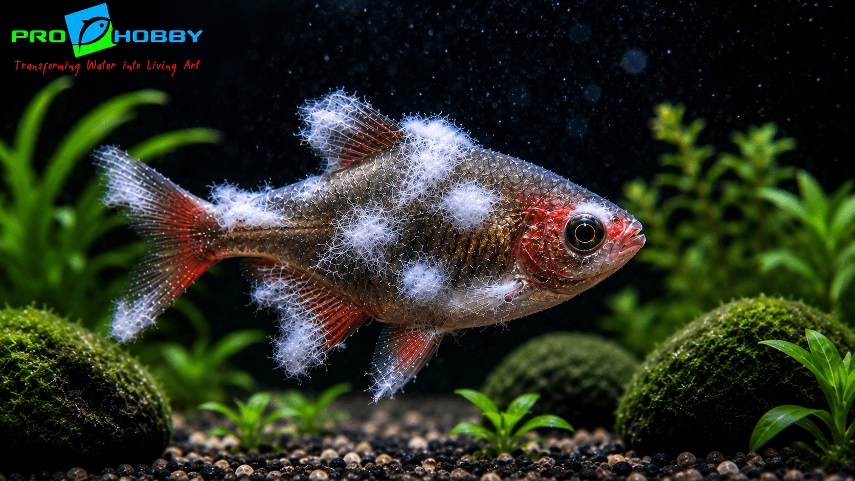
3. What Fungal Infection Looks Like — The Definitive Visual Test
Genuine Saprolegnia infection produces:
- Three-dimensional, fluffy, cotton wool-like tufts extending outward from the infection site into the water
- White, grey, or occasionally brown colouration depending on debris accumulation in the hyphae
- Located specifically at wound sites or areas of prior tissue damage — not spreading uniformly across the body surface
- The growth has genuine depth — it projects away from the fish body
The one-question visual test: Does the white growth extend away from the fish body into the water as a three-dimensional mass? If yes — likely fungal. If the white growth sits flat against the fish surface — it is almost certainly bacterial.
4. Distinguishing Fungal From Bacterial — The Most Critical Differential
This differential diagnosis saves fish lives because antifungals are completely ineffective against bacteria and gram-negative antibiotics have no antifungal activity. Treating the wrong one wastes the critical early treatment window.
| Feature | Saprolegnia (Fungal) | Columnaris (Bacterial) |
|---|---|---|
| Appearance | Fluffy, three-dimensional tufts extending outward | Flat, attached patches on body surface |
| Location | At wound sites specifically | Any body area, fins, mouth |
| Progression | Slower — days to weeks | Fast — can kill in 24–48 hours at warm temperatures |
| Response to antifungal | Effective | None |
| Response to gram-negative antibiotic | None | Effective |
| Treatment | Methylene blue, malachite green, salt | Kanamycin, nitrofurazone |
When in doubt, treat as Columnaris first — it progresses faster and has a narrower treatment window. If gram-negative antibiotics produce no improvement after 3–4 days, reassess and switch to antifungal treatment. The Columnaris Disease guide covers the full bacterial treatment protocol.
Other white conditions to rule out:
- Ich — discrete 1mm spots distributed across body
- Lymphocystis — cauliflower-like growths, viral, no treatment
- External protozoan parasites — grey-blue clouding, not white growth
5. Treatment — Step by Step
Step 1: Isolate in a hospital tank. Prevents antifungal medication from disrupting the main tank’s biological filtration and allows close observation.
Step 2: Identify and address the primary cause. Any underlying wound, bacterial infection, or water quality issue must be treated alongside the fungal infection, not after it.
Step 3: Antifungal treatment — choose one:
Methylene blue — traditional antifungal, particularly effective for egg fungus and mild body infections. 3mg/L in the hospital tank. Warning: stains silicone and equipment permanently blue — use a dedicated hospital tank.
Malachite green — highly effective but toxic to biological filtration and to scaleless fish (loaches, catfish, corydoras). Use in hospital tank only at manufacturer’s recommended dose. Many commercial aquarium fungus products contain malachite green.
Aquarium salt at 2–3g/L — provides osmotic stress on fungal hyphae and supports healing. Effective standalone treatment for mild cases; valuable adjunct for moderate infections.
Step 4: Monitor ammonia daily throughout treatment in the hospital tank. Test with ammonia test kit and manage with small water changes if levels rise.
Step 5: Continue for 7–10 days beyond visible resolution — fungal hyphae can penetrate into tissue below the visible surface.
6. Fungal Infections in Spawning Tanks and Eggs
Saprolegnia is the most significant cause of egg mortality in aquarium breeding. Unfertilised eggs die first and within 24–48 hours develop fungal growth that spreads to adjacent viable eggs, destroying entire spawning batches.
Prevention during breeding:
- Remove unfertilised eggs promptly — they appear white rather than translucent within 24–48 hours of spawning
- Increase water flow over the egg mass — mechanical resistance to fungal colonisation and better oxygenation
- Methylene blue at low concentration (0.1–0.2mg/L) in spawning water provides prophylactic protection without harming eggs or fry
- Maintain optimal water quality — stress on the breeding pair reduces the antifungal compounds they secrete over the egg mass naturally
7. After Treatment — Addressing the Primary Cause
Treating Saprolegnia successfully is only the first step. The primary cause that created the entry point must be resolved to prevent recurrence:
- Identify and remove sharp décor causing physical wounds
- Address any fin-nipping species causing skin damage
- Treat underlying bacterial infections (fin rot, scale rot) that created the bacterial entry point fungus colonised
- Improve water quality — test ammonia, nitrite, and pH. The Complete Water Chemistry Guide covers all parameter interactions.
8. India and Delhi NCR — Specific Considerations
Temperature and Saprolegnia: Saprolegnia thrives in cooler water — it is less prevalent as a spontaneous pathogen in heated tropical aquariums than in temperate conditions. However, physical injury and bacterial infection as primary causes are as common in Indian aquariums as anywhere. The warm Indian summer can increase the speed of secondary fungal colonisation of wounds if fish are already heat-stressed.
Misdiagnosis in the Indian market: Many aquarium shops in India sell general “antifungal” products as a first response to any white growth on fish. The Columnaris misdiagnosis is extremely common — and in a Columnaris case, the antifungal treatment window is being wasted while the fast-moving bacterial disease advances. Aquarium Shop Delhi NCR.
Frequently Asked Questions
My fish has white fluffy growths — is it fungal or bacterial? If the growth is three-dimensional and fluffy, extending away from the fish body at wound sites — likely Saprolegnia fungal infection. If the growth is flat and attached to the body surface, especially not at a specific wound site — almost certainly Columnaris bacterial disease requiring gram-negative antibiotics. Antifungals will not work on Columnaris.
Why isn’t my antifungal medication working? Almost certainly because the condition is bacterial Columnaris, not fungal. The white cottony flat appearance of Columnaris is the consistent source of misdiagnosis. Stop antifungal treatment immediately and switch to gram-negative antibiotics — kanamycin or nitrofurazone. Every day on the wrong treatment is disease progression.
My fish eggs keep developing fungus — how do I prevent this? Remove unfertilised eggs promptly (they turn white within 24–48 hours). Increase water flow over the egg mass. Add methylene blue at 0.1–0.2mg/L to spawning water as prophylaxis. Maintain optimal water quality — the breeding pair’s natural antifungal secretions are compromised under stress.
Can fungal infections spread from fish to fish? Yes — the fungal spores are waterborne and can colonise other fish that have existing wounds or compromised immune function. Isolate affected fish in a hospital tank. Healthy fish with intact skin and good immune function are at low risk even in contaminated water.
Is fungal infection dangerous? Yes if untreated and progressive. Saprolegnia can penetrate into deep tissue, infect internal organs if the infection reaches gill or wound tissue connected to the bloodstream, and the secondary bacterial infection at the fungal colonisation site can develop into septicaemia. Treat promptly.